For learning, not for research. A simplified educational model: the folds it shows are illustrations, not predictions. Scope & limitations
Learn · 1 of 8
Amino acids
Every protein in your body is a chain of just 20 kinds of small molecules, the amino acids, joined end to end. The order of the chain is what makes a hormone a hormone and an enzyme an enzyme.
20standard amino acids are written into the genetic code [1]
9of them are essential: the body cannot make them and they must come from food [1]
~110 Dathe average mass of one amino acid inside a protein [2]
One common frame, 20 different side chains
Every amino acid has the same core: a central carbon (Cα) carrying an amine group (–NH₂), a carboxyl group (–COOH) and a hydrogen. What differs is the fourth attachment, the side chain (R group). Glycine's side chain is a single hydrogen; tryptophan's is a double ring. The side chains decide how each residue behaves: whether it likes or avoids water, whether it carries a charge, whether it can bend, and whether it can form a cross-link.
When two amino acids join, the carboxyl of one reacts with the amine of the next, a water molecule leaves, and a peptide bond (–CO–NH–) is formed. Chains of a few amino acids are peptides; longer chains that fold into a shape are proteins. The repeating –N–Cα–C– backbone is the same everywhere; the side chains stick out from it like charms on a bracelet.
The kinds of amino acids
They are usually grouped by what their side chain does in water. The colours below are the ones the app uses in its Type colour mode.
Gly Glycine · tiny, bends anywhere
Ala Alanine · small, non-polar
Val Valine · non-polar, essential
Leu Leucine · non-polar, essential
Ile Isoleucine · non-polar, essential
Met Methionine · sulfur, the start codon, essential
Pro Proline · ring locks the backbone
Phe Phenylalanine · aromatic ring, essential
Tyr Tyrosine · aromatic, –OH
Trp Tryptophan · double ring, essential
Ser Serine · polar –OH
Thr Threonine · polar –OH, essential
Cys Cysteine · –SH, disulfide bridges
Asn Asparagine · polar amide
Gln Glutamine · polar amide
Lys Lysine · + charge, essential
Arg Arginine · + charge
His Histidine · + at low pH, essential
Asp Aspartate · − charge
Glu Glutamate · − charge
Hydrophobic (water-avoiding)
Gly, Ala, Val, Leu, Ile, Met, Pro and the aromatic Phe, Tyr, Trp. In a folded protein they hide in the core, away from water. This is the main force that folds a protein (see Folding).
Polar, uncharged
Ser, Thr, Cys, Asn, Gln. They like water and hydrogen-bond with it, so they tend to sit on the surface. Two cysteines can bond to each other (a disulfide bridge), stapling a protein together.
Positively charged
Lys, Arg (and His near neutral pH). They attract negative partners, such as DNA, and slow a chain down inside the ribosome's negatively charged exit tunnel.
Negatively charged
Asp, Glu. Together with Lys and Arg they form salt bridges, and they bind positive ions such as Ca²⁺ (calmodulin's EF-hands are loops full of Asp and Glu).
Two special cases: glycine has no side chain, so the backbone can twist freely there; proline's side chain loops back onto the backbone nitrogen, so that joint is stiff and often makes a kink or ends a helix. Both matter a lot for the shape a chain can take [3].
Essential amino acids
Humans can make eleven of the twenty, but not His, Ile, Leu, Lys, Met, Phe, Thr, Trp and Val; these must come from food, and the body recycles them carefully [1]. The app marks them with an e badge.
→
Try it: in the app, hover an amino acid on the left and press Add; the new one floats in, drops a water molecule and bonds to the chain's tail. Add a Gly next to a Pro and compare how freely the chain moves at each joint. Open G-P-G in the app
Sources
Wu G (2009). Amino acids: metabolism, functions, and nutrition. Amino Acids 37:1-17 · DOI
Milo R, Phillips R (2015). Cell Biology by the Numbers. Garland Science, New York · Link
Ramachandran GN, Ramakrishnan C, Sasisekharan V (1963). Stereochemistry of polypeptide chain configurations. J Mol Biol 7:95-99 · DOI
A protein is a chain of amino acids that has folded into a specific shape. The shape is what does the work: it grips, cuts, carries, signals or holds things up.
~20,000protein-coding genes in the human genome [1]
~300amino acids in a typical human protein; the range runs from 9 (oxytocin) to over 30,000 (titin) [2]
~2 × 10⁶ / µm³proteins packed into a cell, a very crowded place [2]
What they are
Chemically a protein is a polymer: one long molecule made of amino acids joined by peptide bonds, usually with a few hundred links. Physically it is a machine part with a shape. The shape comes from the chain folding up on itself, and the sequence of amino acids decides the shape (Anfinsen's principle, see Folding) [3]. Change one amino acid and the shape may hardly change, or the protein may stop working altogether.
Four levels of structure
Primary structure — the sequence itself, written with one letter per amino acid. Insulin's B chain, for example, is FVNQHLCGSHLVEALYLVCGERGFFYTPKT
Secondary structure — local, regular shapes held by hydrogen bonds between backbone N–H and C=O groups: the α-helix (a spiral, 3.6 residues per turn) and the β-sheet (strands lying side by side), both predicted by Pauling in 1951 before anyone had seen one [4]. The app's Protein tab shows a per-residue prediction of these.
Tertiary structure — the full 3D fold of one chain: helices and sheets packed together, with water-avoiding side chains in the middle. Domains are compact, semi-independent parts of a fold (the app lists them under Domains).
Quaternary structure — several chains assembled together. Haemoglobin is four chains; the ATP synthase is dozens [5].
How they work
Almost everything a protein does comes down to shape and stickiness (see Stickiness). A folded protein presents a surface with pockets, grooves and patches of charge. Molecules that fit a pocket bind to it; the rest do not. That is how an enzyme picks its substrate, how an antibody recognises a virus, and how a receptor notices a hormone.
Proteins are not rigid. They breathe, hinge and flip between shapes, and binding one molecule often changes the shape elsewhere, which is how a signal is passed on (allostery). Haemoglobin, the first protein structure solved at this level of detail, changes shape when oxygen binds to one of its four chains, which makes the other three bind more readily [5].
Some proteins have no fixed shape at all over large stretches: intrinsically disordered regions that only fold when they meet a partner. Alpha-synuclein and tau in the app's library are of this kind, which is part of why they are prone to misfolding.
→
Try it: choose Calmodulin in the app, open the Protein tab's Domains and hover an EF-hand, then colour by domain. Open Calmodulin
Sources
UniProt Consortium (2023). UniProt: the Universal Protein Knowledgebase in 2023. Nucleic Acids Res 51:D523-D531 · DOI
Milo R, Phillips R (2015). Cell Biology by the Numbers. Garland Science, New York · Link
Anfinsen CB (1973). Principles that govern the folding of protein chains. Science 181:223-230 · DOI
Pauling L, Corey RB, Branson HR (1951). The structure of proteins: two hydrogen-bonded helical configurations of the polypeptide chain. Proc Natl Acad Sci USA 37:205-211 · DOI
Perutz MF, Rossmann MG, Cullis AF, Muirhead H, Will G, North ACT (1960). Structure of haemoglobin: a three-dimensional Fourier synthesis at 5.5-Å resolution, obtained by X-ray analysis. Nature 185:416-422 · DOI
Proteins are usually sorted by what they do. The same kit of 20 amino acids builds motors, pipes, switches, scaffolds and messengers.
Transport proteins
Carry things. Haemoglobin carries oxygen in the blood; apolipoprotein B (ApoB, 4,536 amino acids) wraps fat into LDL particles; albumin ferries fatty acids and drugs. Membrane transporters and channels move ions and nutrients across cell membranes: the potassium channel lets K⁺ through 10,000 times more readily than the smaller Na⁺, purely by the shape of its pore [1, 2].
Receptors
Sit in the cell membrane and listen. A hormone or neurotransmitter binds on the outside; the receptor changes shape; something happens inside. The largest family, the G-protein-coupled receptors (about 800 in humans), handle light, smell, adrenaline, dopamine and most drugs [3]. The insulin receptor passes its signal on through adaptor proteins such as IRS-1 and IRS-2 (in the library).
Enzymes
Speed up chemical reactions, often a million-fold or more, by holding the reactants in just the right pose. Lysozyme in tears and saliva cuts bacterial cell walls; eNOS makes the nitric oxide that relaxes blood vessels; the aminoacyl-tRNA synthetases load amino acids onto tRNAs for the ribosome.
Hormones & signals
Small proteins and peptides released into the blood or between cells: insulin and glucagon set blood sugar, oxytocin (9 amino acids) triggers labour and bonding. Inside cells, signalling proteins and kinases pass messages on by switching each other on and off.
Structural proteins
Hold things up. Collagen (a quarter of all the protein in the body) forms the ropes of skin, tendon and bone; keratin makes hair and nails; actin and tubulin build the cell's skeleton and tracks.
Motors & machines
Turn chemical energy into movement. Myosin pulls on actin to contract a muscle; kinesin walks along microtubules carrying cargo; the ATP synthase is a rotary motor (see ATP); the ribosome and proteasome are machines for making and breaking proteins.
Defence
Antibodies recognise foreign shapes; complement proteins punch holes in bacteria; glutathione (a 3-residue peptide in the library) and metallothionein mop up reactive molecules.
Storage & regulation
Ferritin stores iron; casein stores amino acids in milk; ubiquitin tags proteins for recycling; transcription factors decide which genes are read.
The app's library sorts its proteins by role (Transport, Hormones, Signalling & regulation, Nervous system, Protection & defence …), the same idea as this page. The one-letter sequences come from UniProt [4].
→
Try it: open the protein picker (Choose) and compare a 9-residue hormone with a 4,536-residue transport protein. Open OxytocinOpen ApoB
Sources
Doyle DA, Morais Cabral J, Pfuetzner RA, Kuo A, Gulbis JM, Cohen SL, Chait BT, MacKinnon R (1998). The structure of the potassium channel: molecular basis of K+ conduction and selectivity. Science 280:69-77 · DOI
Perutz MF, Rossmann MG, Cullis AF, Muirhead H, Will G, North ACT (1960). Structure of haemoglobin: a three-dimensional Fourier synthesis at 5.5-Å resolution, obtained by X-ray analysis. Nature 185:416-422 · DOI
Rosenbaum DM, Rasmussen SGF, Kobilka BK (2009). The structure and function of G-protein-coupled receptors. Nature 459:356-363 · DOI
UniProt Consortium (2023). UniProt: the Universal Protein Knowledgebase in 2023. Nucleic Acids Res 51:D523-D531 · DOI
An adult human makes roughly 4 × 10²¹ protein molecules a day, about 0.3 kg. Each one is read from a gene, copied into messenger RNA and assembled by a ribosome, one amino acid at a time.
1.5 %of the genome codes for proteins: ~20,000 genes [1]
5–6 / samino acids added per second by a human ribosome; bacteria manage ~20 [2, 3]
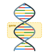
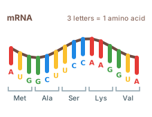
A gene: the recipe for one protein, in the nucleusmRNA: a working copy, three letters per amino acid
The DNA in each cell's nucleus holds a library of recipes (genes) for more than 20,000 different proteins. A gene stores the order of amino acids using four letters (A, C, G, T), three letters per amino acid: a codon. With 64 possible codons for 20 amino acids plus "stop", most amino acids have several spellings; the first codon was decoded in 1961 (UUU = phenylalanine) [4].
When a protein is needed, the gene is copied (transcribed) into a single strand of messenger RNA (mRNA), which is edited, capped and sent out of the nucleus to the ribosomes. Information flows DNA → RNA → protein, the "central dogma" [5]. A single mRNA is usually read many times, by several ribosomes at once (a polysome), before it is broken down.
The ribosome and tRNA
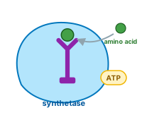
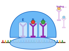
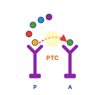
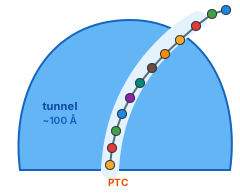
Charging: an enzyme loads each amino acid onto its own tRNA, using ATPThree sites: A (arrive), P (peptide), E (exit)The PTC hands the chain over to the new amino acidThe chain leaves through the exit tunnel, head first
Just in time. Amino acids are not joined straight from the cell fluid. Each is first loaded onto its own transfer RNA (tRNA) by an enzyme, one for each of the 20 amino acids, using the energy of one ATP. The enzyme double-checks and removes most mistakes; a cell holds tens of millions of tRNAs, reloaded over and over.
Three sites. The ribosome holds the mRNA and has three slots for tRNAs: the A site shows the next codon, the P site holds the tRNA carrying the growing chain, and the E site is where empty tRNAs leave. Loaded tRNAs arrive at random and try the A site; wrong ones fall off within milliseconds, so many are tried before one matches. The ribosome picks the wrong amino acid only about once in 10,000.
The bond. At the peptidyl transferase centre (PTC), deep in the large subunit, the amine of the new amino acid attacks the last link of the chain, and the whole chain is handed over to the new amino acid, which becomes the new tail. The PTC is made of RNA, not protein: the ribosome is a ribozyme [6, 7]. Proteins therefore grow from the head (N-terminus) to the tail (C-terminus).
Move on. The ribosome steps one codon along the mRNA, the tRNAs shift A → P → E, and the chain threads further into the exit tunnel: about 100 Å long and 10–20 Å wide, holding some 30–40 amino acids [8]. Positively charged stretches and some sequences touch its walls and slow the ribosome down, giving the chain time to fold [9].
Once the stop codon is reached the chain is released. Many proteins then go through quality control, get chaperone help (see Folding), are modified (sugars, phosphates, cuts) and are carried to where they work; proteins bound for membranes or export are made at the endoplasmic reticulum. If an amino acid runs short, production simply pauses.
→
Try it: choose a protein, press ▶ Build and watch the chain slide out of the ribosome as each new amino acid docks at the PTC; the Translation rate slider sets how many per second. Build the insulin B chain
Sources
UniProt Consortium (2023). UniProt: the Universal Protein Knowledgebase in 2023. Nucleic Acids Res 51:D523-D531 · DOI
Ingolia NT, Lareau LF, Weissman JS (2011). Ribosome profiling of mouse embryonic stem cells reveals the complexity and dynamics of mammalian proteomes. Cell 147:789-802 · DOI
Milo R, Phillips R (2015). Cell Biology by the Numbers. Garland Science, New York · Link
Nirenberg MW, Matthaei JH (1961). The dependence of cell-free protein synthesis in E. coli upon naturally occurring or synthetic polyribonucleotides. Proc Natl Acad Sci USA 47:1588-1602 · DOI
Crick F (1970). Central dogma of molecular biology. Nature 227:561-563 · DOI
Ban N, Nissen P, Hansen J, Moore PB, Steitz TA (2000). The complete atomic structure of the large ribosomal subunit at 2.4 Å resolution. Science 289:905-920 · DOI
Nissen P, Hansen J, Ban N, Moore PB, Steitz TA (2000). The structural basis of ribosome activity in peptide bond synthesis. Science 289:920-930 · DOI
Voss NR, Gerstein M, Steitz TA, Moore PB (2006). The geometry of the ribosomal polypeptide exit tunnel. J Mol Biol 360:893-906 · DOI
Kramer G, Boehringer D, Ban N, Bukau B (2009). The ribosome as a platform for co-translational processing, folding and targeting of newly synthesized proteins. Nat Struct Mol Biol 16:589-597 · DOI
A new chain is a floppy string. Within milliseconds to seconds it folds into the one shape, out of an astronomical number of possibilities, that lets it work. Most proteins start folding while they are still being made.
ms – stypical folding times; a helix turn forms in under a microsecond [1]
~1 kcal/molgained per buried –CH₂– group: small forces, many of them [2]
5–15 kcal/molhow much more stable the folded state is than the unfolded one: only a few hydrogen bonds' worth [2]
The sequence decides the shape
Anfinsen showed in the 1960s that an unfolded enzyme, left alone in water, refolds by itself into its working shape: all the information needed is in the sequence, and the folded state is simply the most stable arrangement [3]. A chain does not search every shape (that would take longer than the age of the universe); it slides down an energy "funnel", forming local structure first and collapsing around its water-avoiding core [1]. Since 2021, deep-learning programs such as AlphaFold can predict most folds from sequence alone [4], which is where the app's Search AlphaFold structures come from.
The forces
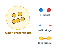
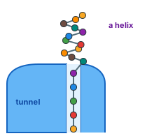
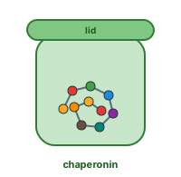
Hydrophobic core, hydrogen bond, salt bridge, disulfide bridgeA helix can already form in the wide end of the exit tunnelA chaperonin gives a difficult protein a private chamber to fold in
The hydrophobic effect is the main driving force: water-avoiding side chains (Leu, Ile, Val, Phe …) gather in the core, and water is happier for it [5, 6].
Hydrogen bonds between backbone N–H and C=O groups make α-helices and β-sheets [7].
Salt bridges between + (Lys, Arg) and − (Asp, Glu) side chains, and van der Waals packing: atoms fitting together tightly like a puzzle.
Disulfide bridges between two cysteines staple a fold shut, mainly in proteins that leave the cell (insulin, antibodies).
Backbone geometry: the peptide bond is flat and stiff, so a chain can only rotate at two joints per residue (φ and ψ), and only some combinations are allowed, the Ramachandran plot [8]. Proline kinks the chain; glycine lets it bend anywhere.
Each of these is weak on its own; a fold holds because hundreds of them act together, and a protein is only marginally stable, so it can still move and be taken apart when its time comes [2].
Folding on the ribosome, and helpers
The exit tunnel is too narrow for most shapes, but a single helix can form in its wider lower part, and small parts fold at its mouth. As more of the chain emerges, domains fold one after another, which helps: the first domain is done before the rest can tangle with it, and slow codons pause the ribosome at difficult spots [9, 10]. At the tunnel exit, chaperones meet the new chain: Hsp70 holds sticky, unfolded stretches so they do not clump, and chaperonins such as TRiC give about one protein in ten a closed chamber to fold in. Chaperones use ATP, but do not dictate the shape; the sequence does [11].
The app's physics has no chaperones (yet): each chain folds on its own, driven by a simplified version of the forces above. See Scope & limitations.
→
Try it: build the insulin B chain, then open the Physics tab and switch the hydrophobic force or the hydrogen bonds off and on; with Fast Physics you can also raise the temperature and watch the fold loosen. Open in the app
Sources
Dill KA, MacCallum JL (2012). The protein-folding problem, 50 years on. Science 338:1042-1046 · DOI
Pace CN, Shirley BA, McNutt M, Gajiwala K (1996). Forces contributing to the conformational stability of proteins. FASEB J 10:75-83 · DOI
Anfinsen CB (1973). Principles that govern the folding of protein chains. Science 181:223-230 · DOI
Jumper J, Evans R, Pritzel A, et al. (2021). Highly accurate protein structure prediction with AlphaFold. Nature 596:583-589 · DOI
Kauzmann W (1959). Some factors in the interpretation of protein denaturation. Adv Protein Chem 14:1-63 · DOI
Chandler D (2005). Interfaces and the driving force of hydrophobic assembly. Nature 437:640-647 · DOI
Pauling L, Corey RB, Branson HR (1951). The structure of proteins: two hydrogen-bonded helical configurations of the polypeptide chain. Proc Natl Acad Sci USA 37:205-211 · DOI
Ramachandran GN, Ramakrishnan C, Sasisekharan V (1963). Stereochemistry of polypeptide chain configurations. J Mol Biol 7:95-99 · DOI
Kramer G, Boehringer D, Ban N, Bukau B (2009). The ribosome as a platform for co-translational processing, folding and targeting of newly synthesized proteins. Nat Struct Mol Biol 16:589-597 · DOI
Balchin D, Hayer-Hartl M, Hartl FU (2016). In vivo aspects of protein folding and quality control. Science 353:aac4354 · DOI
Hartl FU, Bracher A, Hayer-Hartl M (2011). Molecular chaperones in protein folding and proteostasis. Nature 475:324-332 · DOI
Making and moving proteins costs energy, and nearly all of it is paid in one currency: ATP. The protein that mints it is a rotary motor smaller than the wavelength of light.
4 ATPspent per amino acid added: 2 to charge the tRNA, 2 (as GTP) at the ribosome [1]
~ body weightof ATP is made and used every day; each molecule is recycled hundreds of times [1]
120°the rotor's step per ATP: one full turn makes three [2]
ATP
Adenosine triphosphate is a small molecule: an adenine, a sugar and a tail of three phosphates. The phosphates repel each other, so splitting the last one off (to ADP + phosphate) releases energy, about 12 kcal/mol under cell conditions. Almost every process that needs a push, from muscle contraction to pumping ions to building a protein, couples itself to that split. Protein synthesis is the single largest consumer of ATP in a growing cell [1].
ATP synthase
Cells regenerate ATP from ADP mostly in the mitochondria. Burning food pumps protons (H⁺) across the inner mitochondrial membrane, like water behind a dam. The ATP synthase lets them back through, and the flow turns a rotor. The idea that this energy comes from a proton gradient (Mitchell) and that ATP is made by a rotating machine (Boyer's binding-change mechanism) each earned a Nobel prize [3].
Fo, the turbine sits in the membrane. A ring of 8–10 small c-subunits turns as protons pass through, one step per proton [4].
The stalk (γ subunit) is bolted to the ring and spins inside the head.
F1, the head has three catalytic sites arranged around the stalk. As the stalk turns, each site is squeezed through three shapes in turn: bind ADP + phosphate → press them into ATP → open and release it. One full turn makes three ATP [3, 5].
The stator (a second stalk) holds the head still so the rotor can turn against it.
In 1997 the rotation was seen directly: a fluorescent actin filament glued to a single F1 head could be watched spinning under a microscope, in 120° steps, when ATP was supplied, the motor running in reverse [2]. The complete structures, dozens of subunits and thousands of amino acids, have since been resolved by cryo-electron microscopy [4].
The ATP synthase is itself a protein, built and folded like any other: a reminder that the machinery that makes proteins is made of proteins, powered by a protein.
Sources
Milo R, Phillips R (2015). Cell Biology by the Numbers. Garland Science, New York · Link
Noji H, Yasuda R, Yoshida M, Kinosita K (1997). Direct observation of the rotation of F1-ATPase. Nature 386:299-302 · DOI
Boyer PD (1997). The ATP synthase — a splendid molecular machine. Annu Rev Biochem 66:717-749 · DOI
Kühlbrandt W (2019). Structure and mechanisms of F-type ATP synthases. Annu Rev Biochem 88:515-549 · DOI
Walker JE (2013). The ATP synthase: the understood, the uncertain and the unknown. Biochem Soc Trans 41:1-16 · DOI
Folding usually works, but not always. Misfolded proteins are normally refolded or recycled; when they escape and stick together, the result can be a disease.
minutes – yearsthe lifetimes of different proteins before they are recycled [1]
250–300 gof protein broken down and rebuilt by the body every day, three to four times what you eat [2]
> 50human diseases linked to amyloid or misfolded proteins [3]
What goes wrong
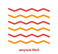
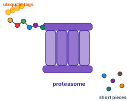
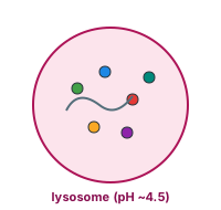
Misfolded chains stacked into an amyloid fibrilTagged with ubiquitin, shredded by the proteasomeBulk digestion in a lysosome (autophagy)Freed amino acids go back into the pool
A chain can fold into the wrong shape, get stuck half-way, or be knocked out of shape later by heat, a mutation, or damage from reactive molecules (the app's RES tab). The common danger is that a misfolded chain exposes the sticky hydrophobic stretches that should be buried in its core. Those stretches find each other, and chains stack into long, very stable fibres called amyloid, held together by β-sheets running the length of the fibre [3, 4].
Many proteins live close to the limit of what the cell can keep soluble, "supersaturated", which is why aggregation shows up so widely in ageing tissue and neurodegeneration [5]. Alzheimer's disease is linked to amyloid-β and tau, Parkinson's to α-synuclein, type 2 diabetes to amylin, and prion diseases to a misfolded prion protein that converts healthy copies on contact. All three brain proteins are in the app's library.
Quality control and recycling
Refold. Chaperones (Hsp70, Hsp90, the chaperonins) catch misfolded chains and give them another try, at the cost of ATP [6].
Tag and shred. Proteins that are old, damaged, misfolded or no longer needed are tagged with a chain of ubiquitin (a 76-residue protein, in the library) and fed into the proteasome, a barrel-shaped machine that unfolds them and cuts them into pieces of 3–25 amino acids [1]. Protein lifetimes range from minutes to years.
Digest in bulk. Larger items, clumps, worn-out organelles and proteins taken in from outside, are wrapped up (autophagy) and digested in lysosomes, acidic compartments full of protein-cutting enzymes.
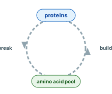
Reuse. Most freed amino acids go back into the pool and into new proteins; the rest are burned, and their nitrogen leaves as urea. Damaged amino acids that resist breakdown build up in cells as lipofuscin, the "age pigment".
With age, chaperones and the proteasome become less effective while damage accumulates, which is why most misfolding diseases are diseases of later life [7].
→
Try it: build Beta-Amyloid (Aβ42) and watch its hydrophobic tail (residues 29–42) collapse; its prefold shows the single chain, not the fibre. Open Aβ42Open Ubiquitin
Sources
Hershko A, Ciechanover A (1998). The ubiquitin system. Annu Rev Biochem 67:425-479 · DOI
Milo R, Phillips R (2015). Cell Biology by the Numbers. Garland Science, New York · Link
Chiti F, Dobson CM (2017). Protein misfolding, amyloid formation, and human disease: a summary of progress over the last decade. Annu Rev Biochem 86:27-68 · DOI
Knowles TPJ, Vendruscolo M, Dobson CM (2014). The amyloid state and its association with protein misfolding diseases. Nat Rev Mol Cell Biol 15:384-396 · DOI
Ciryam P, Tartaglia GG, Morimoto RI, Dobson CM, Vendruscolo M (2013). Widespread aggregation and neurodegenerative diseases are associated with supersaturated proteins. Cell Rep 5:781-790 · DOI
Hartl FU, Bracher A, Hayer-Hartl M (2011). Molecular chaperones in protein folding and proteostasis. Nature 475:324-332 · DOI
Balchin D, Hayer-Hartl M, Hartl FU (2016). In vivo aspects of protein folding and quality control. Science 353:aac4354 · DOI
Proteins work by sticking to things, and go wrong by sticking to the wrong things. What makes a surface sticky is mostly water.
Why hydrophobic means sticky
An oily side chain does not attract anything strongly; the trouble is that water around it has to arrange itself into an ordered cage, which water "dislikes" (it costs entropy). Put two oily patches together and the cage shrinks: water is released and the patches stay stuck. This hydrophobic effect is what folds a protein, and it is also what makes any exposed hydrophobic patch on a protein's surface sticky [1, 2]. The same force makes oil droplets merge and lipid membranes form.
Good stickiness
Binding partners. Enzymes grip substrates, antibodies grip antigens, receptors grip hormones, all through patches of matching shape and charge. Most binding surfaces mix a hydrophobic centre with charged or polar rims that set the orientation.
Assemblies. Haemoglobin's four chains, the ribosome's ~80 proteins and the ATP synthase's rotor are all held together by sticky interfaces.
In the tunnel. The ribosome exit tunnel has charged walls; positively charged stretches (Lys, Arg) cling to them and slow translation down, which the app reproduces with its wall electrostatics [3].
Bad stickiness
A cell is extremely crowded: proteins fill 20–30 % of its volume and bump into each other constantly. Evolution therefore keeps protein surfaces about as non-sticky as it can; across species, the more abundant a protein, the fewer sticky residues it shows on its surface, because a surplus of chance contacts would gum up the cell [4]. Misfolding breaks this rule by exposing hydrophobic core stretches, and the result is aggregation and amyloid (see Misfolding) [5]. Chaperones exist largely to cover sticky stretches until they can be buried [6].
Stickiness is also a dial. A moderately sticky protein can form reversible droplets (membrane-less organelles) that concentrate reactions; too sticky, and the droplets harden into fibres. Cells tune this with phosphorylation and with the charge of their surroundings.
In the app
The physics libraries model stickiness as a pairwise attraction between hydrophobic side chains (the Hydrophobic force), electrostatics between charged ones, and a Stickiness setting in the Build group that lets hydrophobic residues drag on the walls near the tunnel exit. Prefolds and single-chain builds cannot show two chains sticking together: there is only ever one chain.
→
Try it: build Melittin (bee venom, 26 residues), an amphipathic helix with one oily face and one charged face, then set the colour mode to Type to see the two faces. Open Melittin
Sources
Kauzmann W (1959). Some factors in the interpretation of protein denaturation. Adv Protein Chem 14:1-63 · DOI
Chandler D (2005). Interfaces and the driving force of hydrophobic assembly. Nature 437:640-647 · DOI
Kramer G, Boehringer D, Ban N, Bukau B (2009). The ribosome as a platform for co-translational processing, folding and targeting of newly synthesized proteins. Nat Struct Mol Biol 16:589-597 · DOI
Levy ED, De S, Teichmann SA (2012). Cellular crowding imposes global constraints on the chemistry and evolution of proteomes. Proc Natl Acad Sci USA 109:20461-20466 · DOI
Knowles TPJ, Vendruscolo M, Dobson CM (2014). The amyloid state and its association with protein misfolding diseases. Nat Rev Mol Cell Biol 15:384-396 · DOI
Hartl FU, Bracher A, Hayer-Hartl M (2011). Molecular chaperones in protein folding and proteostasis. Nature 475:324-332 · DOI